- Startseite
- blinker birne
- X-ray manual : U.S. Army . <*5 lit w Fig. 3. Position for first exposure.. Fig. 4. Position for second exposure.200 HEAD EXAMINATIONS 201 moved from the front plane of the
X-ray manual : U.S. Army . <*5 lit w Fig. 3. Position for first exposure.. Fig. 4. Position for second exposure.200 HEAD EXAMINATIONS 201 moved from the front plane of the
5 (247) · € 32.00 · Auf Lager
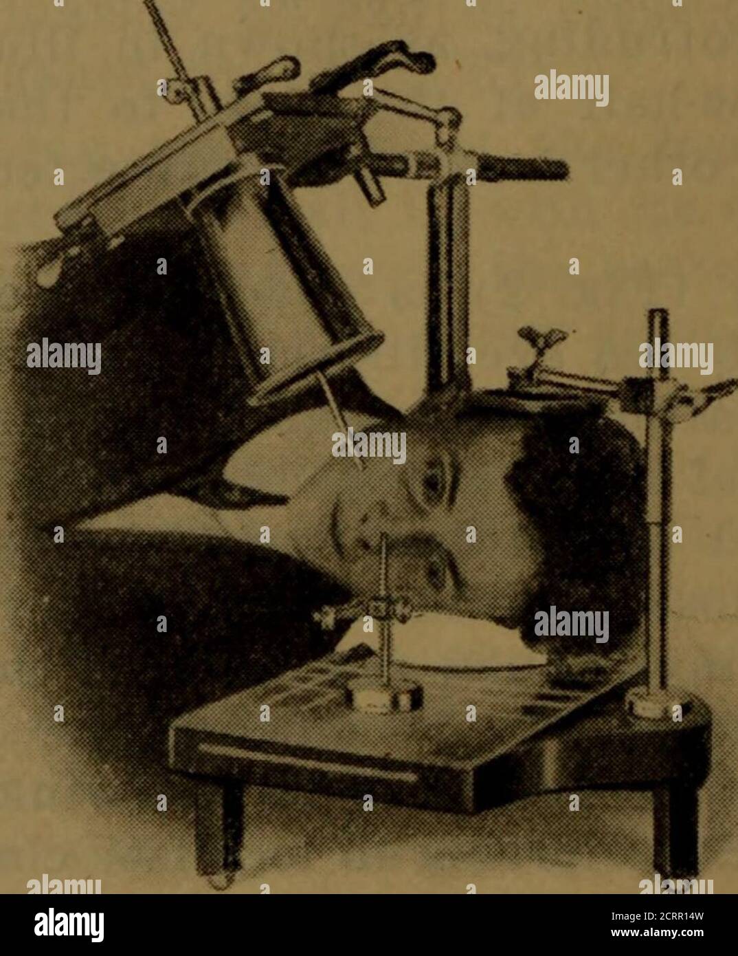
Download this stock image: . X-ray manual : U.S. Army . <*5 lit w Fig. 3. Position for first exposure.. Fig. 4. Position for second exposure.200 HEAD EXAMINATIONS 201 moved from the front plane of the cornea, and it shouldalso be borne in mind that the front of the cornea is 10millimeters in front of the shadow of the indicator-ball, asshown in your negatives. The tube is now centered overthe localizing ball and cone so that the shadows of thetwo will coincide (Fig. 3). Some object, such as a candle or a piece of whitepaper, that can be readily seen by the patient, should beplaced in alignment with the sights of the - 2CRR14W from Alamy's library of millions of high resolution stock photos, illustrations and vectors.

Howitzer tm9 319 by Veteran Corps of Artillery of the State of New York - Issuu

Radiology - ScienceDirect

Principles of Naval Weapons Systems by Francisco Azevedo - Issuu

Miscellaneous - Clark's Positioning In Radiography - by A. S. Whitley

IJSPT V17N1: The Human Movement System by IJSPT - Issuu

phywe-tess-phy-lep-en by SIDLAB,S. L. - Issuu

IJSPT Volume 17 Number 2 by IJSPT - Issuu

Distance from the magnification device contributes to differences in lower leg length measured in patients with TSF correction
Diffi hi-res stock photography and images - Page 13 - Alamy

IJSPT Volume 17, Number 3 by IJSPT - Issuu

First x ray tube hi-res stock photography and images - Alamy